Pityriasis Amiantacea: The Asbestos Scalp Condition Most People Have Never Heard Of.
Pityriasis Amiantacea:
The Asbestos Scalp Condition
Most People Have Never Heard Of.
Thick, silvery scales binding hair shafts into matted tufts. Alarming but usually temporary hair loss. Frequently mistaken for severe dandruff. This is the complete clinical picture: what it is, what drives it, how clinicians identify it, and what actually helps.
Pityriasis amiantacea is a sign of an underlying medical condition, and identifying that condition requires a clinician. This article is educational, not medical advice. Kapyderm products are cosmetic scalp care formulas — they are not drugs, not antifungals, and not a treatment for PA or anything driving it. PA can be caused by a fungal infection (tinea capitis), which needs antifungal medicine, and heavy scaling with hair loss should always be assessed by a dermatologist. Never stop a prescribed medication on your own. Individual results vary.
Pityriasis amiantacea (also called tinea amiantacea, asbestos scalp, or pseudotinea amiantacea) is a reactive scalp condition where thick, asbestos-like scales encircle hair shafts and bind them in matted tufts. It is not a primary diagnosis but a reaction pattern driven by an underlying condition. In the largest published series — 85 patients at a single Egyptian hospital — scalp psoriasis was confirmed in 35.3% of cases and seborrheic dermatitis in 34.2%, and the authors stress keeping tinea capitis in mind when evaluating PA.[1] Standard anti-dandruff shampoos generally do not shift it, because the scale is anchored around the hair shaft rather than sitting loose on the surface. Hair loss is usually transitory. Getting the driver identified is the whole job, and that is a clinician's, not a product's.
- What pityriasis amiantacea actually is — history and mechanism
- Who gets it — what the published series show
- The underlying causes — what drives the PA reaction pattern
- Drug-induced pityriasis amiantacea
- Recognizing it — signs and symptoms
- Why anti-dandruff shampoos fail
- Diagnosis — trichoscopy and clinical examination
- Differential diagnosis — PA vs dandruff vs psoriasis vs seborrheic
- The hair loss question — what to expect
- Treatment options — honest comparison
- The Dermotricology plant-based routine
- The evidence behind the botanical routine
- Frequently asked questions
What Pityriasis Amiantacea Actually Is — History and Mechanism
The name has a specific etymology worth knowing: pityriasis comes from the Greek pityron (bran or scaling), and amiantacea from the French amiante — asbestos — because the thick, silvery, layered scales reminded early clinicians of asbestos fiber. It was first described by French dermatologist Jean-Louis-Marc Alibert in his 1832 Monographie des dermatoses.[2] It appears in the literature under several alternate names: tinea amiantacea, pseudotinea amiantacea, and asbestos scalp.
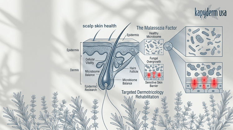
The mechanism is structural, not just surface scaling. Inflammation in the scalp drives an exaggerated keratinocyte proliferation and cohesion response. Rather than shedding normally, dead skin cells accumulate in a densely packed, layered structure that infiltrates the space around the proximal hair shaft near the follicle opening. Multiple shafts become encased in this scale shell — bound in tufts with a characteristic overlapping "roof tile" pattern. Histopathology in the 85-patient series confirmed diffuse hyperkeratosis and parakeratosis in every biopsy taken.[1]
Pityriasis amiantacea is a reaction pattern, not a primary diagnosis — the authors of the largest study state this as their central conclusion.[1] It is always driven by an underlying inflammatory or infectious condition. That is why softening the scale without identifying the driver leads to recurrence — and why the first step for anyone with this presentation is a clinical assessment, not a product.
Who Gets It — What the Published Series Show
Those percentages come from one prospective study of 85 patients at a single dermatology department in Mansoura, Egypt, collected in 2000–01.[1] It remains the largest series published, and it is genuinely the best evidence available — but it is one hospital, one population, one two-year window. Most articles quote these figures as universal rates. They are the best estimate we have, not a global average. A separate Korean series of 44 cases found a wider range of underlying conditions, including pemphigus vulgaris, lichen planopilaris and folliculitis decalvans alongside the usual drivers.[3]
PA is reported more often in women, and it occurs at any age, including children — where tinea capitis features more prominently as a driver.[9] It is generally considered uncommon, though underdiagnosis is likely given how often it is mistaken for severe dandruff.
The Underlying Causes — What Drives the PA Reaction Pattern
Because PA is a secondary reaction pattern, identifying the underlying driver is the single most important clinical step — it determines the treatment entirely. The same external presentation can arise from very different root causes, and only one of the routes below is a fungal infection needing antifungal medicine.
Scalp Psoriasis
IL-23/Th17 immune dysregulation accelerates skin cell turnover, and the hyperproliferative response produces the thick scale that triggers the PA pattern. Where psoriasis is the driver, trichoscopy tends to show twisted red loops and dotted vessels beneath the scale.
Seborrheic Dermatitis & Atopic Eczema
Malassezia-associated inflammation or immune-mediated barrier dysfunction creates the scalp environment behind the PA scale response. Seborrheic-driven PA typically produces yellower scales; atopic-driven PA may show more redness in the surrounding skin.
Tinea Capitis (Fungal Infection)
A scalp fungal infection, most commonly Trichophyton or Microsporum. Particularly relevant in children. The authors of the largest study specifically stress keeping tinea capitis in mind when evaluating PA.[1] It needs prescription antifungal treatment — no cosmetic product treats it.
Lichen Simplex Chronicus
A chronic itch-scratch cycle producing localized thickening and reactive scaling that can escalate to the PA pattern. Breaking the itch cycle matters as much as addressing the scale.
Scarring Alopecias
The Korean series identified lichen planopilaris and folliculitis decalvans among underlying conditions.[3] These are scarring conditions where hair loss can be permanent — a reason PA with hair loss belongs in front of a dermatologist rather than being self-managed.
Idiopathic PA
When no underlying condition is found despite thorough evaluation, PA is classified as idiopathic — as in the Mayo Clinic case report, where Wood lamp and fungal culture were used to exclude tinea capitis first.[4]
Drug-Induced Pityriasis Amiantacea
One of the least-known aspects of pityriasis amiantacea is its documented association with pharmaceutical triggers — an angle almost entirely absent from consumer-facing content.
TNF-alpha inhibitor therapy
PA has been reported as a distinctive presentation of psoriasis associated with TNF-alpha inhibitor therapy — the class of biologics used for psoriasis, rheumatoid arthritis, Crohn's disease and ankylosing spondylitis — in Clinical and Experimental Dermatology in 2012.[5] It is a paradoxical association, since these drugs are themselves used to treat psoriasis. If you are on biologic therapy and develop PA, raise it with your rheumatologist or dermatologist.
Valproic acid
A case report in the International Journal of Trichology documented PA developing after the start of valproic acid — an anticonvulsant used for epilepsy, bipolar disorder and migraine prevention.[6] A single case report is the weakest form of published evidence, and it is included here because it is what exists, not because the link is established.
If PA appeared or significantly worsened after starting a new medication — particularly a biologic or an anticonvulsant — that timing is worth raising with the prescribing physician. It does not mean the medication should be stopped, and it must not be stopped on your own. It means the conversation is a medical one.
Recognizing It — Signs and Symptoms
The hallmark features are specific enough that experienced clinicians often identify PA on visual examination. The following signs collectively distinguish it from other scaling scalp conditions:
- Thick, adherent silvery or yellow scales — unlike dandruff flakes that fall freely, PA scales cling tightly to both scalp and hair shaft and resist brushing and standard shampooing. Silver-white is more typical of psoriatic-driven PA; yellow of seborrheic-driven PA.
- The "roof tile" or sheath pattern — scales overlap in a layered structure encircling the proximal hair shaft. This is the most diagnostically specific feature and is best seen on trichoscopy.[8]
- Hair shaft tufting and matting — multiple shafts bound within the same scale crust. Pulling tufts apart without first softening the scale causes breakage.
- Itching — commonly the most prominent symptom, though severity varies widely between individuals.
- Patchy distribution — PA can affect any area of the scalp, often following the underlying condition's own distribution.
- Usually non-scarring hair loss in affected areas — the Mayo Clinic case documented non-scarring hair loss in a woman in her early 30s with frontal and temporal involvement.[4] "Usually" is doing real work in that sentence — see the hair loss section below.
Why Anti-Dandruff Shampoos Fail Against PA
This is one of the most common points of confusion — and a common reason people arrive at a consultation having tried every anti-dandruff product available without result.
Standard anti-dandruff shampoos are formulated to address surface-level flaking. Their actives work by reducing Malassezia populations, slowing surface cell turnover, or loosening surface scale cohesion.
PA scales are not loose surface flakes. They are anchored to the proximal hair shaft in a dense, layered crust that a brief wash-and-rinse cycle generally cannot penetrate. The scale encases the hair shaft, not just the scalp surface.
Softening that scale takes a keratolytic left in contact with the area, applied repeatedly over time rather than in a single wash. Emollient oils under occlusion are a well-established way to do this. But softening scale is only half the problem — and the less important half. The underlying condition still has to be identified and treated, and that is what determines whether the scale comes back.
Diagnosis — Trichoscopy and Clinical Examination
Diagnosis is a clinical act, performed by a physician. The distinctive visual presentation is usually sufficient for an experienced clinician, and trichoscopy adds precision — a 2012 study in Anais Brasileiros de Dermatologia established the dermatoscopic features of the scale-encrusted hair tufts characteristic of PA, alongside microscopy of the tufts themselves.[8]
The vascular pattern visible beneath the scale can point toward the underlying driver — twisted red loops and dotted vessels toward psoriasis, arborizing vessels toward seborrheic dermatitis. That is a clinician's inference to draw, and it is confirmed by examination and testing, not by pattern-matching alone.
When fungal culture and Wood lamp are essential
Where tinea capitis is suspected — particularly in children — fungal culture and Wood lamp examination should be performed before treatment begins. The Mayo Clinic case report used exactly this sequence to exclude tinea capitis before classifying the case as idiopathic.[4] Treating fungal PA without antifungal coverage risks treatment failure and spread to others.
Certified Kapyderm centers use the Kapykon camera to look at the scalp surface at magnification. That is a cosmetic evaluation — it is not a diagnosis, and a Technician of Dermotricology is not a substitute for a dermatologist. Its useful role here is recognizing when a scalp needs medical referral, and adapting a gentle care routine around whatever the physician has diagnosed.
Differential Diagnosis — PA vs Dandruff vs Scalp Psoriasis vs Seborrheic
| Feature | Dandruff (mild SD) | Scalp Psoriasis | Seborrheic Dermatitis | Pityriasis Amiantacea |
|---|---|---|---|---|
| Scale appearance | White/grey, loose, fine | Silver-white, thick plaques | Yellow-greasy, loosely adherent | Silver/yellow, very thick, layered — adheres to shaft |
| Scale location | Scalp surface only | Scalp surface and plaque | Scalp surface, oily zones | Encircles and binds to the hair shaft — "roof tile" pattern |
| Hair shaft binding | None | None (scale on scalp) | None | Yes — the defining feature. Multiple shafts bound in tufts |
| Hair matting | No | No | No | Yes — tufts matted at the root |
| Response to standard shampoo | Improves | Partial improvement | Improves with antifungal | Little response — scale anchored to the shaft |
| Trichoscopy vascular pattern | Normal or mild arborizing | Twisted red loops, dotted vessels | Arborizing vessels | Reflects whichever condition is driving it |
| Hair loss | None | Mild if severe | Rare | Usually transitory; scarring rare but documented |
| What treatment targets | Surface microbiome | IL-23/Th17 pathway | Malassezia and inflammation | The underlying driver first; scale softening second |
The Hair Loss Question — What to Expect
This is the most emotionally significant aspect of PA for most people, and the aspect most inadequately addressed by existing content. Clear counseling belongs before treatment begins, not after someone notices hair in the sink and panics.
Hair loss in PA is commonly transitory, and scarring alopecia is described as extremely rare in the largest series.[1] That is the reassuring and accurate general picture. But it is not never: a 1991 paper in Acta Dermato-Venereologica reported PA specifically as an unrecognized cause of scarring alopecia in four patients,[7] and the Korean series found lichen planopilaris and folliculitis decalvans — both scarring conditions — among underlying diagnoses.[3] Most articles quote the reassurance and omit the caveat. Both belong here, because the caveat is the reason to be seen by a dermatologist rather than wait it out.
What is generally understood about PA-related shedding:
- Hair that comes away during scale removal is often hair that was trapped within the crust, released as the scale is softened
- In non-scarring PA the follicle itself is intact, and hair regrows once the underlying condition is treated
- Forcibly pulling scale off without softening it first causes more breakage than gradual softening does
- The regrowth timeline depends on the underlying condition and how long it went untreated
See a dermatologist promptly if hair loss is accompanied by scalp scarring, smooth shiny areas, or a visible absence of follicular openings. Those signs suggest a scarring alopecia, where hair loss can be permanent and where time matters.
Treatment Options — Honest Comparison
Conventional PA treatment follows a standard dermatological approach: soften and remove the scale, then treat the underlying condition. Both halves are needed.
Coal tar preparations
A long-standing first-line option. Coal tar slows cell turnover and loosens scale. The practical drawbacks are well known: it stains, has a strong persistent odor, causes photosensitivity, and there is a long-running safety discussion in the literature about its polycyclic aromatic hydrocarbon content. The clinical consensus is that therapeutic coal tar at regulated concentrations is acceptable for its intended use; the real-world problem is that people struggle to keep using it.
Topical corticosteroids
Commonly prescribed to suppress the underlying inflammatory driver, particularly where psoriasis is confirmed. Effective, and subject to the usual long-term considerations — tachyphylaxis, skin atrophy with prolonged use, and rebound on abrupt discontinuation. They do not soften existing scale, so they work best once the surface has been cleared.
Antifungals (ketoconazole, ciclopirox, or oral agents)
Essential where Malassezia or tinea capitis is the driver. Confirmed tinea capitis generally requires an oral antifungal — topical treatment alone is usually inadequate. This is prescription territory and needs a physician.
Emollient soaking (oils)
Vegetable or mineral oils applied under occlusion for several hours soften the scale enough to allow gentle removal. Genuinely useful as a preparatory step, inexpensive, and low-risk. Not therapeutic on its own — it does not address the underlying condition or prevent recurrence.
The Dermotricology Plant-Based Routine
What follows is a cosmetic scalp care routine — gentle cleansing and daily comfort care for a scalp that is scaling. It is not a treatment for PA or for anything causing it, and it does not replace the medical assessment described above. People often use a routine like this alongside what their dermatologist has prescribed. Tell your dermatologist what you are using, and patch test first.
These statements have not been evaluated by the Food and Drug Administration. This product is not intended to diagnose, treat, cure, or prevent any disease. Food supplements do not replace a varied diet or prescribed medication. Tell your physician about every supplement you take, particularly if you are on medication for a scalp or autoimmune condition.
How quickly scale softens depends far more on the underlying condition and the medical treatment for it than on any cosmetic routine, so we would rather not put a number on it. Some hair coming away as scale releases is common and is not the same as follicle damage. If scaling is not improving, or hair loss continues, that is a reason to go back to the dermatologist rather than to persist with a scalp routine.
The Evidence Behind the Botanical Routine
Below is what the independent literature says about the mechanisms this routine works through — and, just as importantly, what it does not say.
Well established
The prospective study of 85 PA patients found scalp psoriasis in 35.3% and seborrheic dermatitis in 34.2%, and concluded PA is a particular reaction pattern of the scalp to various inflammatory scalp diseases.[1] This is the best-supported claim on this page, and it is the one that matters most clinically — because it means the product question is secondary to the diagnosis question.
Supported in laboratory study
The Depure supplement is led by green nettle (Urtica dioica), with dandelion, boldo, fumitory and peppermint. Nettle-leaf polyphenol fractions have been applied to human skin cells under oxidative and inflammatory stress with measurable antioxidant and anti-inflammatory activity, and the characterized actives are the phenolic acids chlorogenic, caffeic and p-coumaric acid.[10] The limit: that is isolated plant fractions applied to cells in a dish — not a capsule, not a person, and not PA.
An active research area, not settled
The gut-skin axis — the link between intestinal microbiome balance and inflammatory skin disease — is a genuine and active field, with reviews examining its role in psoriasis and atopic dermatitis.[11] The limit, stated plainly: association is not causation, none of this work involves pityriasis amiantacea, and no study has tested a Kapyderm supplement in any of these conditions.
What the literature does not show
These studies concern mechanisms and ingredients, not this routine. No controlled trial of any Kapyderm product in pityriasis amiantacea exists, and no independent study has compared it with conventional care. This is a cosmetic scalp care routine, and PA is a sign of an underlying medical condition that needs medical assessment.
Frequently Asked Questions
- Abdel-Hamid IA, Agha SA, Moustafa YM, El-Labban AM. Pityriasis amiantacea: a clinical and etiopathologic study of 85 patients. Int J Dermatol. 2003;42(4):260–264. doi:10.1046/j.1365-4362.2003.01755.x. PMID 12694489. Prospective study at a single department, Mansoura Faculty of Medicine, Egypt, 2000–01. Scalp psoriasis 35.3%; seborrheic dermatitis 34.2%; staphylococci present in 96.5%; hyperkeratosis and parakeratosis in all biopsies; authors conclude PA is a reaction pattern and stress keeping tinea capitis in mind. pubmed
- Alibert JL. Monographie des dermatoses, ou précis théorique et pratique des maladies de la peau. Paris: Daynac; 1832:293–295. The original description.
- Kwak HB, Yun SK, Kim HU, Park J. Pityriasis amiantacea: an epidemiologic study of 44 cases in Korean patients. Ann Dermatol. PMID 33911716. Retrospective series; underlying conditions included seborrheic dermatitis, psoriasis, pemphigus vulgaris, lichen planopilaris, folliculitis decalvans and tinea capitis. pubmed
- Xie F, Sominidi-Damodaran S, et al. Pityriasis Amiantacea. Mayo Clinic Proceedings. 2022. doi:10.1016/j.mayocp.2022.06.003. Woman in her early 30s; frontal and temporal scalp; non-scarring hair loss on presentation; Wood lamp and fungal culture used to exclude tinea capitis; classified as idiopathic. mayoclinicproceedings.org
- Pityriasis amiantacea: a distinctive presentation of psoriasis associated with tumour necrosis factor-α inhibitor therapy. Clin Exp Dermatol. 2012;37(6):639–641. The source of the TNF-alpha inhibitor association.
- Diaz-Perez JA, Joyce JC, Cibull TL, Victor TA. Development of pityriasis amiantacea after valproic acid therapy. Int J Trichology. PMID 30607045. A single case report — the weakest form of published evidence. pubmed
- Langtry JAA, Ive FA. Pityriasis amiantacea, an unrecognized cause of scarring alopecia, described in four patients. Acta Derm Venereol (Stockh). 1991;71:352–353. The counterpoint to the "scarring is rare" picture.
- Verardino GC, Azulay-Abulafia L, Macedo PM, Jeunon T. Pityriasis amiantacea: clinical-dermatoscopic features and microscopy of hair tufts. An Bras Dermatol. 2012;87(1):142–145. The trichoscopic reference for the scale-encrusted hair tuft.
- DermNet NZ. Pityriasis amiantacea. Clinical reference resource — reaction pattern rather than primary diagnosis; reported female predominance; occurs in children and young adults. dermnetnz.org
- Salachna P, et al. Oxidative stress protection and anti-inflammatory activity of a polyphenolic fraction from Urtica dioica: in-vitro study using human skin cells. 2024. Nettle-leaf polyphenols showed antioxidant and anti-inflammatory activity in human skin cells under stress. In-vitro only.
- Gut-skin axis reviews: The gut-skin axis: a bidirectional, microbiota-driven relationship with therapeutic potential. Gut Microbes. 2025;17(1):2473524. · Chen M, Wang R, Wang T. Gut microbiota and skin pathologies: mechanism of the gut-skin axis in atopic dermatitis and psoriasis. Int Immunopharmacol. 2024;141:112658. Neither concerns pityriasis amiantacea.
11 sources: 8 peer-reviewed journal articles, 1 historical monograph, 1 clinical reference resource, and 1 pair of review articles. Refs 6 and 7 are case reports and small series — named as such rather than presented as established fact. No controlled trial of any Kapyderm product in pityriasis amiantacea exists. The percentages in this article come from a single-centre Egyptian study and are the best available evidence, not a global average — this article says so rather than quoting them as universal. Last reviewed August 2026.
Gentle Daily Scalp Care.
Alongside Your Dermatologist, Not Instead of One.
The Kapyderm scalp care range is a plant-based cosmetic system for daily comfort on a scaling, sensitive scalp — gentle cleansing and barrier support. EU-regulated. If you have pityriasis amiantacea, get the underlying cause identified first: that is the part that determines the outcome.